

❤️ Unit-9: Transport in Animals
Cambridge IGCSE Biology (0610)
❤️ Topic Area: Human Biology
⏱️ Estimated Reading Time: 30-35 minutes
🎯 Difficulty Level: ★★★★☆
📖 Last Updated: October 2026
Understand how the circulatory system transports substances around the body, how the heart and blood vessels function, how blood components support transport and defence, and how cardiovascular health can be maintained.
🎯 Learning Objectives
By the end of this unit, you should be able to:
- 1. Describe the structure and function of the circulatory system, including single and double circulation.
- 2. Identify and explain the structure of the mammalian heart, the pathway of blood flow, and the role of valves in maintaining one-way circulation.
- 3. Compare the structure and functions of arteries, veins and capillaries, relating their adaptations to their roles in transport.
- 4. Identify and explain the components of blood and their functions in oxygen transport, defence, clotting and the transportation of substances.
- 5. Explain the mechanism of blood clotting and the roles of platelets, fibrinogen and fibrin in preventing blood loss and pathogen entry.
- 6. Investigate and explain the effect of physical activity on heart rate and understand the factors that influence cardiovascular health.
- 7. Describe and evaluate the causes, risk factors and preventive measures of coronary heart disease, including the roles of diet and exercise.
📋 Syllabus Inclusion
Tick each point when you feel confident that you have learned it.
1.1 The Circulatory System
Most animals have a circulatory system that transports substances around the body. In mammals, the circulatory system consists of:
- Blood: transports substances such as oxygen, nutrients, carbon dioxide and other waste products.
- Blood vessels: form a network of tubes through which blood flows.
- Heart: acts as a pump that generates pressure to circulate blood.
- Valves: ensure that blood flows in one direction and prevent backflow.
The circulatory system transports useful substances to cells and carries waste products away from them.
Why is a circulatory system necessary?
In small organisms, substances may move directly between cells and the surrounding environment by diffusion.
However, mammals have a large body size and a relatively small surface area compared with their volume. Diffusion alone would be too slow to supply all cells with oxygen and nutrients and remove waste products.
Therefore, a circulatory system is needed for efficient transport throughout the body.
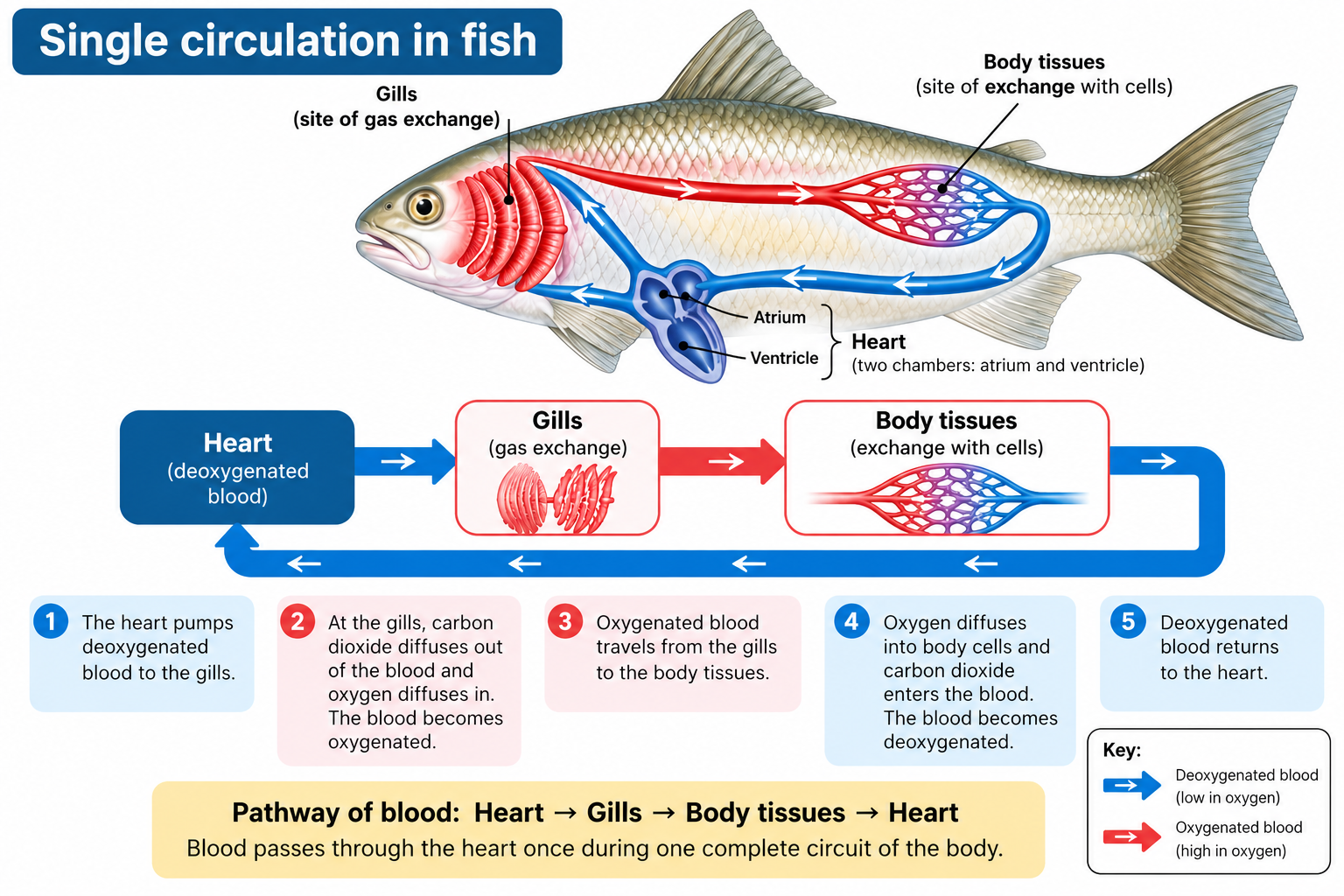
1.2 Single Circulation in Fish:
Single circulation is a type of circulation in which blood passes through the heart once during one complete circuit of the body.
Fish have a single circulatory system.
Pathway of blood in fish: Heart → Gills → Body tissues → Heart
The process occurs as follows:
- Blood is pumped from the heart to the gills.
- At the gills, carbon dioxide diffuses out of the blood and oxygen diffuses into it.
- The blood becomes oxygenated.
- Oxygenated blood travels from the gills to the body tissues.
- Oxygen diffuses from the blood into body cells.
- Blood becomes deoxygenated and returns to the heart.
Thus, blood passes through the heart only once during a complete circuit.
Key feature
In fish, blood pressure decreases as blood passes through the narrow capillaries of the gills. Therefore, blood reaches the body tissues at relatively low pressure.
1.3 Double Circulation in Mammals:
What is double circulation?
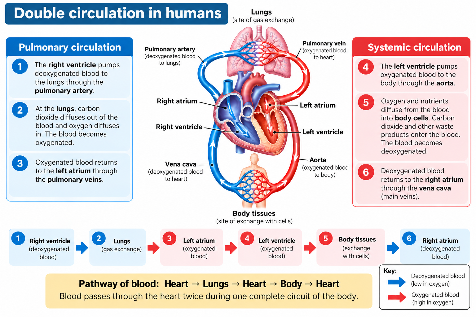
Double circulation is a type of circulation in which blood passes through the heart twice during one complete circuit of the body.
Mammals have a double circulatory system consisting of two circuits:
- Pulmonary circulation
- Systemic circulation
The mammalian heart is divided into right and left halves by a muscular wall called the septum. Each half performs a different role in circulation.
A. Pulmonary circulation
Pulmonary circulation is the movement of blood between the heart and the lungs.
- The right side of the heart pumps deoxygenated blood to the lungs.
- In the lungs, carbon dioxide diffuses from the blood into the air spaces, while oxygen diffuses into the blood.
- The blood becomes oxygenated.
- Oxygenated blood returns to the left side of the heart.
B. Systemic circulation
Systemic circulation is the movement of blood between the heart and the rest of the body.
- The left side of the heart pumps oxygenated blood to the body.
- Oxygen and nutrients are delivered to body cells.
- Carbon dioxide and other waste products enter the blood.
- Blood becomes deoxygenated and returns to the right side of the heart.
Complete pathway of double circulation
The complete route is: Heart → Lungs → Heart → Body → Heart
The blood passes through the heart twice:
- Once during pulmonary circulation.
- Once during systemic circulation.
Advantages of Double Circulation
Double circulation has two important advantages.
Advantage 1: Different blood pressures can be maintained
The right side of the heart pumps blood to the lungs at relatively low pressure.
- This prevents damage to the delicate capillaries in the lungs.
- It allows sufficient time for gas exchange to occur.
The left side of the heart pumps blood to the rest of the body at much higher pressure.
- Blood travels over a much greater distance.
- It must overcome greater resistance to flow.
- Higher pressure allows oxygen and nutrients to be delivered rapidly to body tissues.
Advantage 2: Efficient oxygen supply
Double circulation ensures that oxygenated blood returns to the heart before being pumped to the body.
This allows blood to be pumped to the body at high pressure, providing an efficient supply of oxygen for aerobic respiration.
This is particularly important in mammals, which have high energy requirements.
Comparison: Single and Double Circulation
| Feature | Single circulation | Double circulation |
| Example | Fish | Mammals |
| Times blood passes through heart in one complete circuit | Once | Twice |
| Number of circuits | One | Two |
| Main pathway | Heart → Gills → Body → Heart | Heart → Lungs → Heart → Body → Heart |
| Blood pressure to body | Relatively low | High |
| Separation of pulmonary and systemic circuits | No separate circuits | Two distinct circuits |
Key Terms to Remember:
| Term | Meaning |
| Circulatory system | System of blood vessels with a pump and valves to ensure one-way flow of blood |
| Single circulation | Blood passes through the heart once in one complete circuit |
| Double circulation | Blood passes through the heart twice in one complete circuit |
| Pulmonary circulation | Blood flow between the heart and lungs |
| Systemic circulation | Blood flow between the heart and the rest of the body |
| Oxygenated blood | Blood containing a high concentration of oxygen |
| Deoxygenated blood | Blood containing a lower concentration of oxygen |
2.1 The Heart as a Pump
- The heart is a muscular organ that pumps blood around the body.
- It works as two pumps operating side by side:
- The right side pumps deoxygenated blood to the lungs.
- The left side pumps oxygenated blood to the rest of the body.
- The heart therefore maintains the two circuits of the mammalian double circulatory system.
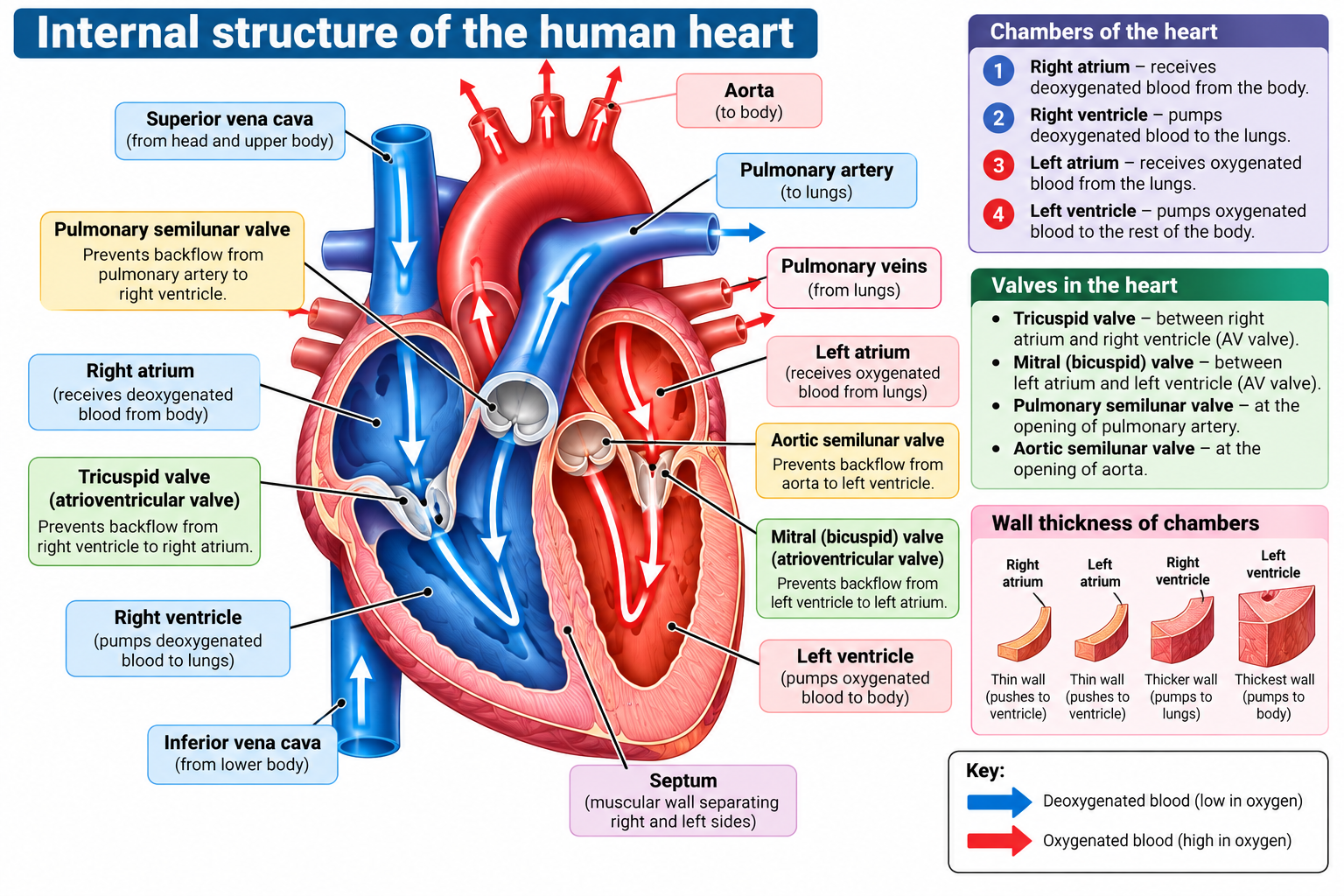
- The mammalian heart has four chambers:
- Right atrium: The right atrium receives deoxygenated blood returning from the body. It passes this blood into the right ventricle.
- Right ventricle: The right ventricle pumps deoxygenated blood to the lungs. This forms the beginning of pulmonary circulation.
- Left atrium: The left atrium receives oxygenated blood returning from the lungs. It passes this blood into the left ventricle.
- Left ventricle: The left ventricle pumps oxygenated blood to the rest of the body. This begins systemic circulation.
- The two upper chambers are the atria, while the two lower chambers are the ventricles.
- Atria receives the blood while ventricles pump out the blood.
2.2 The Septum and The Valves:
The septum is a muscular wall that separates the right and left sides of the heart. Its importance is that it prevents the mixing of oxygenated and deoxygenated blood.
This allows:
- the right side to handle deoxygenated blood;
- the left side to handle oxygenated blood;
- oxygenated blood to be delivered efficiently to body tissues.
The heart contains one-way valves. Their function is to prevent blood from flowing backwards. There are two groups of valves to understand at this level:
A) Atrioventricular valves
- These are located between: the atria and ventricles.
- They allow blood to move from atria to the ventricles but prevent it from flowing back into the atria when the ventricles contract.
- The AV valve between left atria and left ventricle is “Mitral or Bicuspid Valve” while the one between right atria and right ventricle is “Tricuspid Valve”.
B) Semilunar valves
- These are located at the opening of the pulmonary artery in the right ventricle and the opening of the aorta in the left ventricle.
- They prevent blood from flowing back into the ventricles after it has been pumped out.
2.3 Relative Thickness of Heart Walls
All the chambers of the heart do not have equally thick muscular walls. The walls of the atria are thinner than the walls of the ventricles. Why?
- The atria only need to push blood a short distance into the ventricles.
- The ventricles need to pump blood out of the heart.
- Therefore, the ventricles require more muscular walls.
Also, the walls of ventricles are not equally thick. The left ventricle has a much thicker muscular wall than the right ventricle. Why?
- The left ventricle pumps blood around the whole body
- The right ventricle only pumps blood to the lungs.
Therefore, longer the distance to be travelled, higher the pressure required to be pushed. So to exert more pressure, more thicker walls are needed. This relationship between wall thickness and the distance/pressure over which blood must be pumped is an important concept.
2.4 The Cardiac Cycle:
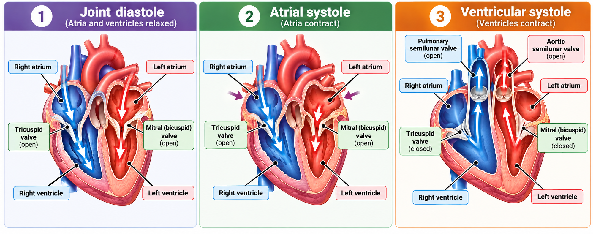
The cardiac cycle is the sequence of events that occurs during one complete heartbeat. It can be described in three main steps:
Step 1: Joint Diastole — Heart relaxes
- The atria and ventricles are relaxed.
- Blood flows into the atria from the vena cava and pulmonary veins.
- The atrioventricular valves are open, so blood flows from the atria into the ventricles.
- The semilunar valves are closed, preventing blood from flowing back into the ventricles.
- The ventricles gradually fill with blood.
Step 2: Atrial Systole — Atria contract
- The atria contract.
- This pushes the remaining blood in the atria into the ventricles.
- The atrioventricular valves remain open.
- The semilunar valves remain closed.
- The ventricles become completely filled with blood.
Step 3: Ventricular Systole — Ventricles contract
- The ventricles contract.
- Pressure inside the ventricles rises.
- The atrioventricular valves close, preventing blood from flowing back into the atria.
- The semilunar valves open.
- Blood is forced out:
- Right ventricle → pulmonary artery → lungs
- Left ventricle → aorta → body
- The atria are relaxed and begin filling with blood again.
2.5 Structure–Function Relationships:
| Structure | Function / Advantage |
| Right atrium | Receives deoxygenated blood from the body |
| Right ventricle | Pumps deoxygenated blood to the lungs |
| Left atrium | Receives oxygenated blood from the lungs |
| Left ventricle | Pumps oxygenated blood to the body |
| Septum | Separates oxygenated and deoxygenated blood |
| Valves | Prevent backflow and maintain one-way flow |
| Atria | Have thinner walls because they only push blood into ventricles |
| Ventricles | Have thicker walls because they pump blood out of the heart |
| Left ventricle | Thickest wall because it pumps blood around the whole body |
| Right ventricle | Less thick wall because it pumps blood only to the lungs |
| Coronary arteries | Supply the heart muscle with blood |
3.1 Components of Blood
Blood is composed of two components; a liquid component called plasma and a cellular component.
The cellular component has three main types of cells; Red blood cells (Erythrocytes), White blood cells (Leucocytes), and Platelets (Thrombocytes)
A) Liquid Component: Plasma
Plasma is the liquid part of blood.
It is mainly water and contains dissolved substances.
Plasma transports:
- blood cells;
- ions;
- nutrients;
- urea;
- hormones;
- carbon dioxide.
Therefore, plasma provides the liquid medium through which many substances are transported around the body.
B) Cellular Component: RBC, WBC, and Platelets
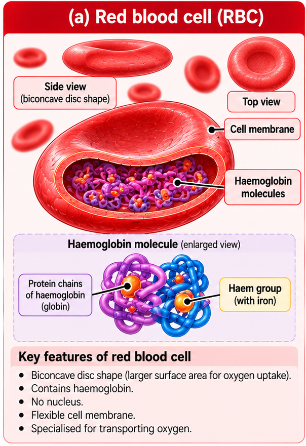
a) Red Blood Cells (RBC)
Red blood cells are specialised for transporting oxygen.
Red blood cells:
- contain protein haemoglobin;
- have a large surface area compared with their volume;
- have no nucleus;
- have a biconcave disc shape.
Haemoglobin
Haemoglobin is a protein containing iron.
Haemoglobin + oxygen ⇌ oxyhaemoglobin
Oxygen is therefore loaded onto haemoglobin.
At body tissues, oxygen is released from oxyhaemoglobin and diffuses into cells.
Function
The main function of red blood cells is:
Transport of oxygen from the lungs to body tissues.
b) White Blood Cells (WBC)
White blood cells defend the body against disease.
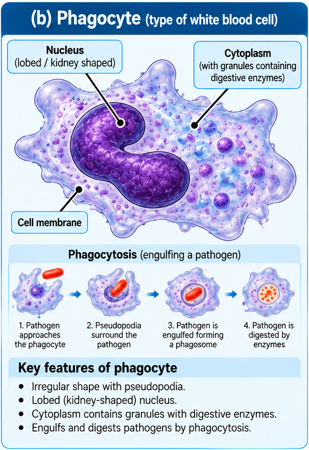
1) Phagocytes
Phagocytes defend the body by engulfing and digesting pathogens.
This process is called phagocytosis.
The basic sequence is: Pathogen encountered → phagocyte surrounds pathogen → pathogen is engulfed → pathogen is digested
Phagocytes can move towards areas where pathogens are present and engulf them.
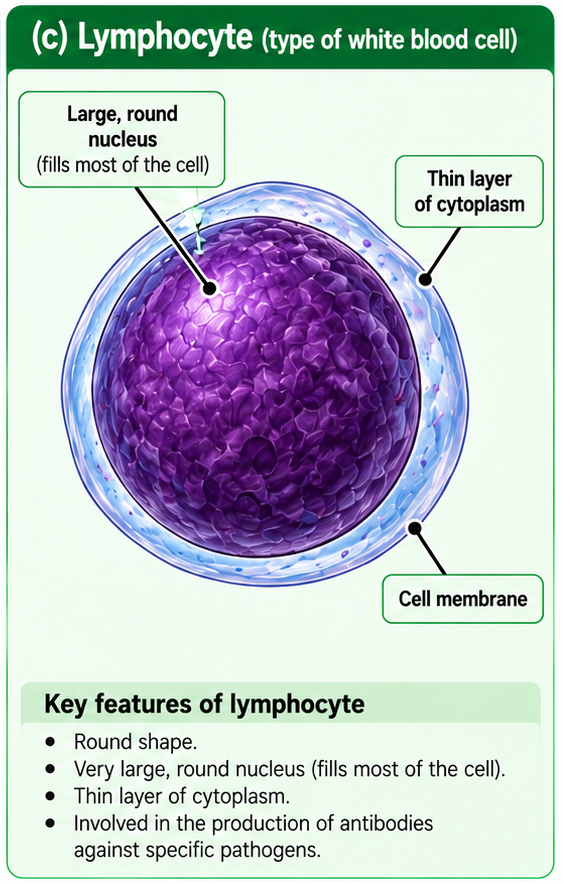
2) Lymphocytes
Lymphocytes are involved in producing antibodies.
When a pathogen enters the body, lymphocytes produce antibodies that are specific to the pathogen.
The antibody binds to the corresponding antigen on the pathogen.
This contributes to the body’s defence against disease.
See the details of role of phagocytes and lymphocytes in providing the body’s immune response in Unit-10 Diseases and Immunity
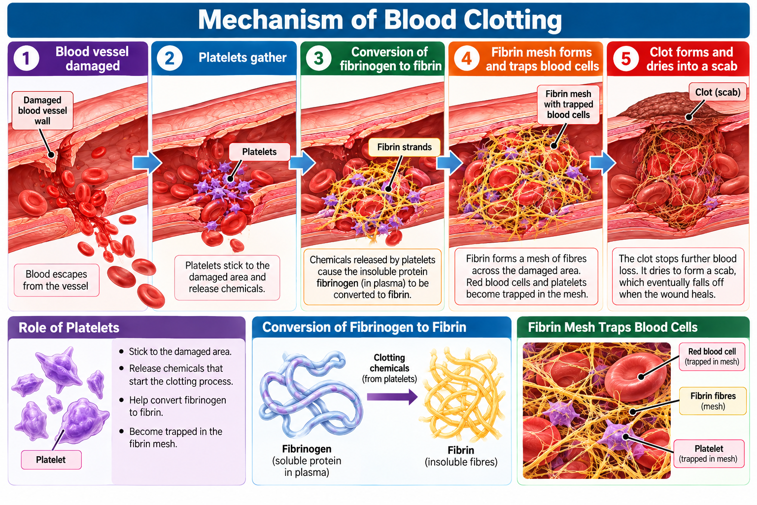
c) Platelets
Platelets are small fragments involved in blood clotting.
They are important when a blood vessel is damaged.
Their main role is to help form a clot that:
- prevents excessive blood loss;
- helps prevent pathogens from entering the body.
3.2 Blood Vessels
Blood vessels are tubes that transport blood around the body. Each type has a structure suited to its function. There are three main types:
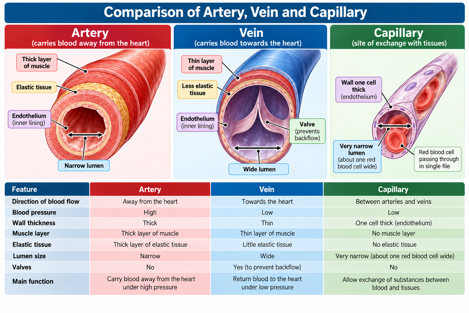
1. Arteries:
Structure:
- Thick muscular walls
- Elastic tissue in their walls
- A relatively small/narrow lumen
- No valves along their length
Function
Arteries carry blood away from the heart under high pressure.
The heart’s contractions produce pressure that pushes blood into the arteries.
Why are artery walls thick and muscular?
The blood entering arteries is at high pressure.
The thick muscular and elastic walls:
- withstand the high pressure;
- prevent the vessel from bursting;
- allow the artery to maintain its shape.
Why is elastic tissue important?
During the ventricular contraction, blood is pushed into the arteries with high pressure. To withstand this pressure the artery wall stretches slightly.
When the heart relaxes, the elastic tissue recoils.
This helps maintain blood pressure and keeps blood flowing through the artery.
2. Veins
Structure
- Thinner walls than arteries
- Less muscle and elastic tissue than arteries
- A larger lumen than arteries
- Presence of Valves
Function
Veins return blood to the heart.
The pressure of blood in veins is much lower than in arteries.
Why do veins have valves?
Because blood pressure is low, blood could flow backwards, particularly in veins carrying blood upwards against gravity.
Valves ensure that blood flows in one direction towards the heart.
When blood tries to flow backwards, the valves close.
3. Capillaries
Capillaries are extremely small blood vessels that connect the smallest arteries to the smallest veins.
Capillaries have:
- Walls only one cell thick
- A very narrow lumen
- No thick muscular or elastic wall
Function
Capillaries are the main sites where substances are exchanged between the blood and body tissues.
They allow movement of substances between blood and surrounding tissues, such as:
- oxygen;
- nutrients;
- carbon dioxide;
- other dissolved substances
Why are capillaries only one cell thick?
The thin wall provides a short diffusion distance, allowing substances to move efficiently between the blood and surrounding cells.
Why are capillaries numerous?
The body contains a very extensive network of capillaries. This provides a large surface area for exchange and ensures that most cells are close to a blood supply.
Comparison of Blood Vessels
| Feature | Artery | Vein | Capillary |
| Direction of blood flow | Away from heart | Towards heart | Between arteries and veins |
| Pressure | High | Low | Low |
| Wall | Thick | Thin | One cell thick |
| Muscle and elastic tissue | Thick layer | Thinner layer | Absent |
| Lumen | Relatively narrow | Wide | Very narrow |
| Valves | No | Yes | No |
| Main function | Carry blood away from heart | Return blood to heart | Exchange substances with tissues |
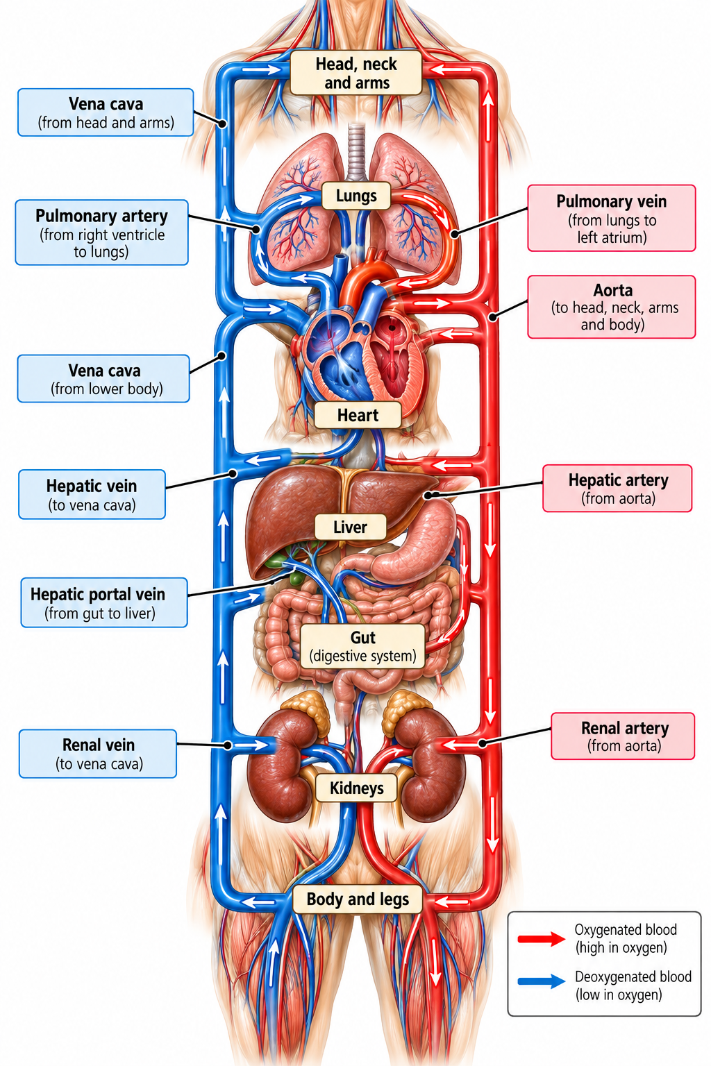
3.3 Main Blood Vessels
Blood vessels associated with the heart
- Aorta – It is the largest artery. It originates from the left ventricle and carries oxygenated blood away from the heart. It divides into multiple arteries and supplies oxygen rich blood to all the organs and tissues of the body.
- Vena cava – It is the largest vein. It carries blood from the upper part of the body (superior vena cava) and lower part of the body (inferior vena cava) and returns deoxygenated blood from the body to the right atrium.
- Pulmonary artery – It originates from the right ventricle. After coming out from the heart, the pulmonary artery divides into two main branches and carries deoxygenated blood to each (left and right) lungs.
- Pulmonary vein – It originates from the lungs and carries oxygenated blood from the lungs to the left atrium.
- Coronary artery – It branches from the aorta and supplies oxygenated blood to the heart muscle (cardiac muscles). The oxygen is used in aerobic respiration to release energy required for continuous contraction of the heart.
Blood vessels associated with the lungs
- Pulmonary artery – Carries deoxygenated blood from the right ventricle to the lungs.
- Pulmonary vein – Carries oxygenated blood from the lungs to the left atrium.
Blood vessels associated with the kidneys
- Renal artery – It branches from the aorta and carries oxygenated as well as unfiltered (urea rich) blood to the kidney.
- Renal vein – It carries deoxygenated as well as filtered (urea poor) blood away from the kidney and joins the inferior vena cava.
Blood vessels associated with the liver:
- Hepatic artery – It branches from the aorta and carries oxygenated blood to the liver. The liver uses this oxygen for aerobic respiration.
- Hepatic vein – It carries blood away from the liver to the inferior vena cava. This blood contains substances processed by the liver and has a lower oxygen concentration than the blood entering through the hepatic artery.
- Hepatic portal vein – It carries blood rich in absorbed nutrients from the gut to the liver. The liver processes, stores or converts these nutrients as required by the body.
This section explores how coronary heart disease affects the functioning of the heart, how physical activity influences heart rate, and how lifestyle choices can help reduce the risk of cardiovascular disease.
4.1 Coronary Heart Disease (CHD)
Structure and function of coronary arteries
The heart is a muscular organ that requires a continuous supply of oxygen and nutrients to carry out its contractions. Although blood passes through the chambers of the heart, the heart muscle receives its own blood supply through the coronary arteries.
The coronary arteries branch from the aorta and carry oxygenated blood to the heart muscle. This blood supplies oxygen and glucose needed for aerobic respiration, which releases energy for cardiac muscle contraction.
What is coronary heart disease?
Coronary heart disease (CHD) occurs when the coronary arteries become narrowed or blocked, reducing the supply of blood and oxygen to the heart muscle.
The narrowing commonly occurs when fatty deposits build up in the inner walls of the arteries. These deposits can form plaques that reduce the diameter of the lumen, restricting blood flow. If a plaque ruptures, a blood clot may form and further obstruct the artery.
The reduced blood supply means that the heart muscle receives less oxygen for aerobic respiration. Consequently, less energy may be available for normal contraction.
- Angina: If the oxygen supply to the heart muscle is insufficient, a person may experience chest pain or discomfort, particularly during physical exertion.
- Heart attack: If a coronary artery becomes severely or completely blocked, part of the heart muscle may receive too little oxygen and become damaged or die.
The severity of the effects depends on the extent and duration of the blockage and the area of heart muscle affected.
4.2 The Heart and Exercise
Effect of physical activity on heart rate
Heart rate is the number of times the heart beats per minute (bpm). It changes according to the body’s requirements for oxygen and energy.
During physical activity, skeletal muscles contract more frequently and require more energy. The rate of aerobic respiration increases, so muscle cells need a greater supply of oxygen and glucose. They also produce carbon dioxide more rapidly.
The heart responds by beating faster, increasing the volume of blood pumped per minute. This helps deliver oxygen and glucose to working muscles and transport carbon dioxide away from them.
Changes in heart rate during and after exercise
- At rest: The heart beats at a rate sufficient to meet the body’s normal energy requirements.
- During exercise: Heart rate increases as the muscles require more oxygen and glucose for respiration. The increase depends on the intensity and duration of the activity.
- Immediately after exercise: Heart rate remains elevated because the body continues to meet the increased demands associated with recovery.
- During recovery: Heart rate gradually decreases towards its resting level as the muscles’ energy requirements return to normal.
The time taken for heart rate to return towards its resting level varies with the individual, their fitness, and the intensity of exercise.
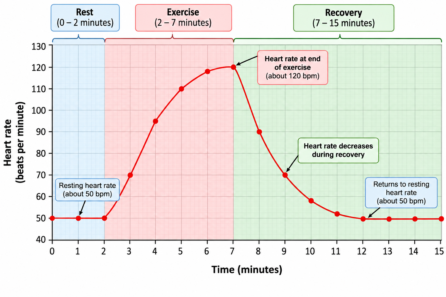
Investigating the effect of exercise on heart rate
The effect of physical activity on heart rate can be investigated by measuring the pulse before and after exercise.
1. Measure resting heart rate: Allow the participant to rest quietly, then count their pulse for one minute.
2. Perform physical activity: Ask the participant to perform a defined exercise for a fixed duration.
3. Record heart rate: Measure heart rate immediately after exercise and at regular intervals during recovery.
4. Analyse the results: Compare the readings and plot heart rate against time to identify the increase during exercise and the decrease during recovery. For a fair comparison, the exercise duration, intensity, measurement method and recovery intervals should be kept consistent. Repeating the investigation and calculating mean values can improve reliability.
Methods of monitoring heart activity
The activity of the heart can be monitored in three main ways.
a) Electrocardiogram (ECG)
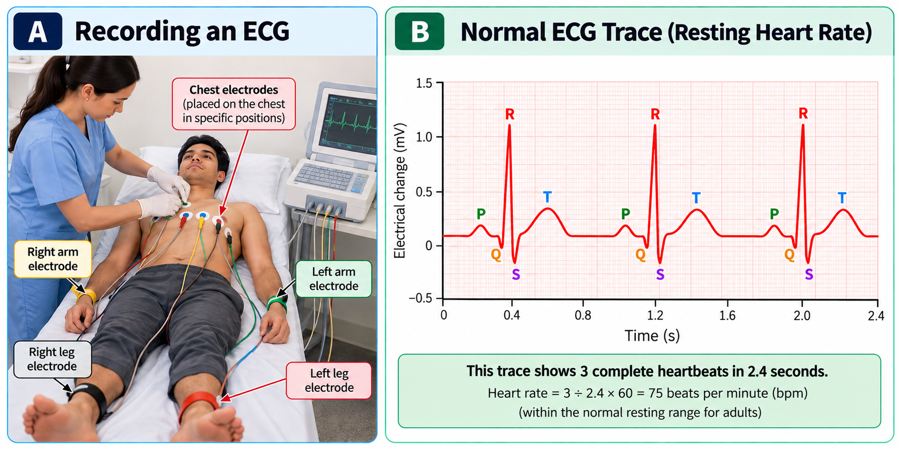
An ECG records the electrical activity of the heart. Electrodes are placed at different positions on the body and the trace can be used to assess heart rate and the pattern of electrical activity during each heartbeat on a monitor screen.The EGC graph is read with reference to waves represented as P, Q, R and S. This represents different stages of electrical activity associated with the contraction of the heart chambers.
| Wave | What it represents | What happens in the heart |
| P wave | Atrial depolarisation | Electrical activity spreads through the atria, triggering atrial contraction (atrial systole). |
| Q wave | Beginning of ventricular depolarisation | Electrical activity begins spreading through the ventricles. |
| R wave | Main part of ventricular depolarisation | Electrical activity spreads rapidly through much of the ventricular muscle. |
| S wave | Final part of ventricular depolarisation | Electrical activity spreads through the remaining ventricular muscle. |
An important distinction: Depolarisation is the electrical event; contraction is the mechanical response. The ECG records electrical activity, not the contraction itself.
b) Pulse rate
Each heartbeat produces a pressure wave that can be felt in an artery. Pulse rate can be measured at places where an artery passes close to the surface of the skin and can be pressed gently against an underlying bone. Counting these pulses per minute provides an estimate of heart rate.
Common sites for measuring pulse rate
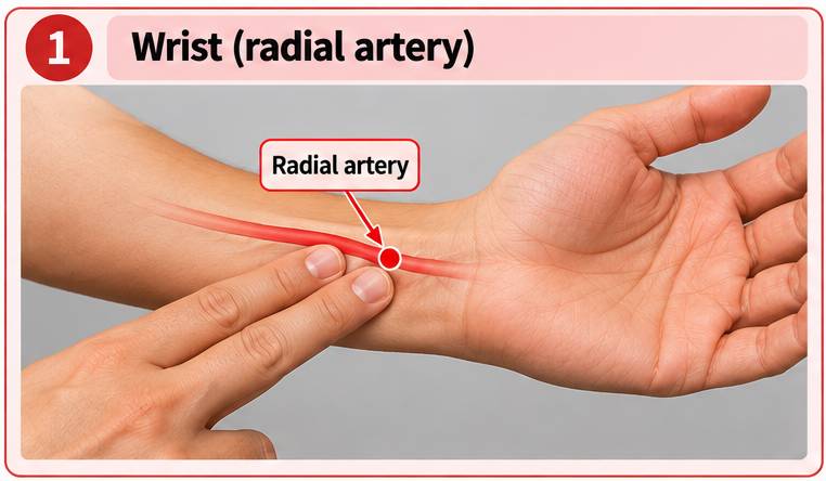
1. Wrist (radial artery): The most common site. Place two fingers on the thumb side of the wrist.
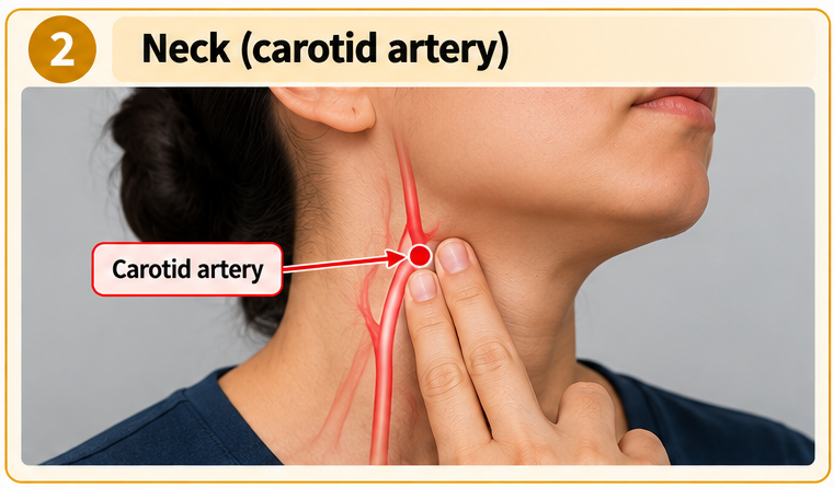
2. Neck (carotid artery): The pulse can be felt on either side of the neck, beside the windpipe. Press gently and measure on one side only.
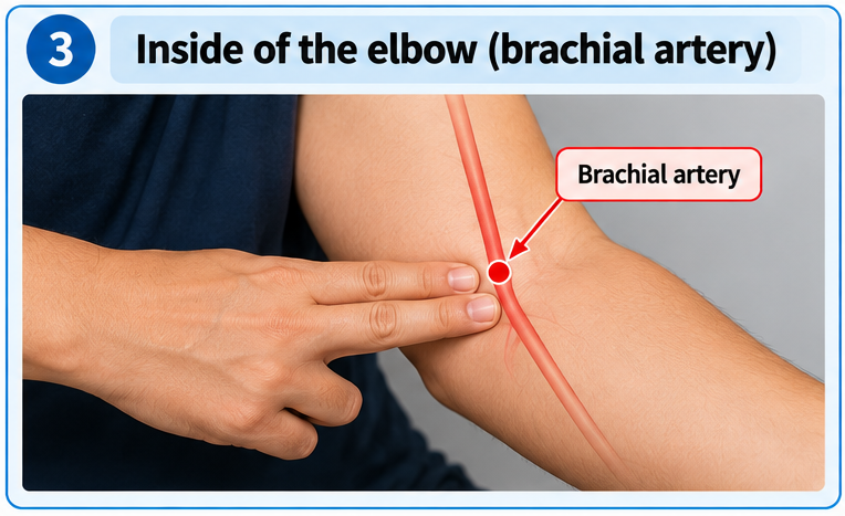
3. Inside the elbow (brachial artery): The pulse can be felt on the inner side of the elbow, particularly during certain clinical measurements.
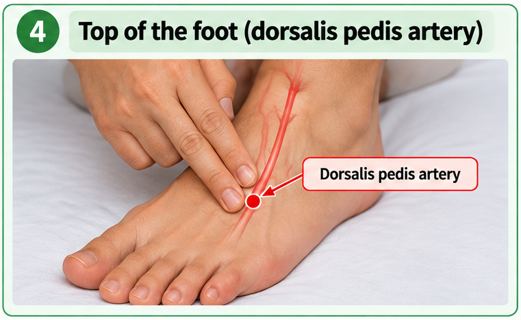
4. Top of the foot (dorsalis pedis artery): The pulse can be felt on the upper surface of the foot.
Important: Use the index and middle fingers, not the thumb, because the thumb has its own detectable pulse.
c) Listening to heart-valve sounds
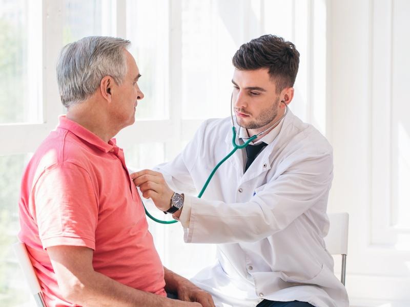
A stethoscope allows the sounds produced mainly by the closing of the heart valves to be heard. These sounds provide information about the timing of events in the cardiac cycle.
First heart sound — “Lub”
During ventricular contraction, pressure inside the ventricles rises. This causes the atrioventricular valves to close, preventing blood from flowing back into the atria. Their closure produces the first heart sound.
Second heart sound — “Dub”
During ventricular relaxation, pressure in the arteries causes the semilunar valves to close. This prevents blood from flowing backwards into the ventricles and produces the second heart sound.
Heart rate, anaerobic respiration and recovery
During vigorous exercise, the muscles may not receive oxygen quickly enough to meet all their energy requirements through aerobic respiration. Anaerobic respiration then releases additional energy, producing lactic acid in human muscle cells.
After exercise, heart rate and breathing rate may remain elevated. This helps transport lactic acid in the blood from the muscles to the liver and supply oxygen for its aerobic respiration. The additional oxygen required to deal with the lactic acid is associated with the oxygen debt.
The continued increase in heart rate after exercise is therefore part of the body’s recovery process, rather than an immediate return to resting conditions.
4.3 Risk Factors for Coronary Heart Disease
A risk factor is a characteristic or behaviour that increases the likelihood of developing a disease. Several factors can increase the risk of coronary heart disease. Some can be modified through lifestyle changes, while others cannot.
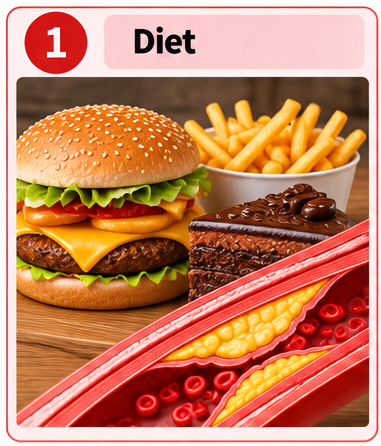
1. Diet: A diet high in saturated fat and trans-fat can contribute to the build-up of fatty deposits in artery walls. Excessive salt intake can contribute to high blood pressure, placing additional strain on the cardiovascular system.
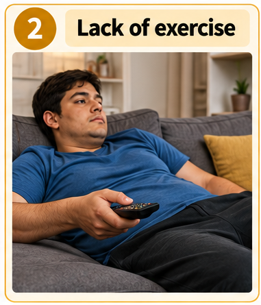
2. Lack of exercise: Physical inactivity increases the risk of poor cardiovascular fitness and can contribute to excess body fat, high blood pressure and unhealthy blood lipid levels.
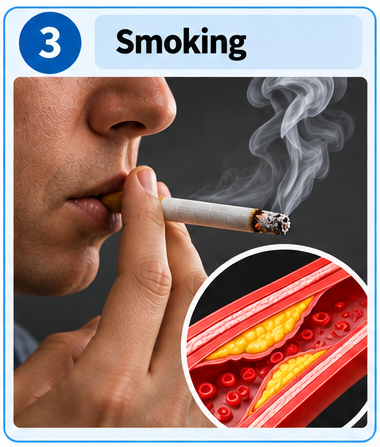
3. Smoking: Chemicals in tobacco smoke damage blood vessel linings. Nicotine increases heart rate and blood pressure, while carbon monoxide reduces the oxygen-carrying capacity of blood. Smoking also promotes processes that contribute to atherosclerosis and blood clot formation.
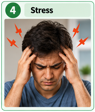
4. Stress: Persistent stress can contribute to elevated blood pressure and may encourage behaviours that increase cardiovascular risk, such as smoking, physical inactivity or unhealthy eating.
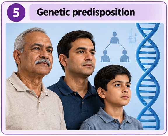
5. Genetic predisposition: Inherited characteristics can influence a person’s susceptibility to CHD, including their tendency to develop high blood pressure or abnormal blood cholesterol levels.
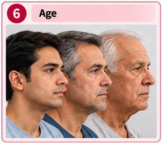
6. Age: The risk of CHD generally increases with age because fatty deposits and damage to blood vessels can accumulate over time.
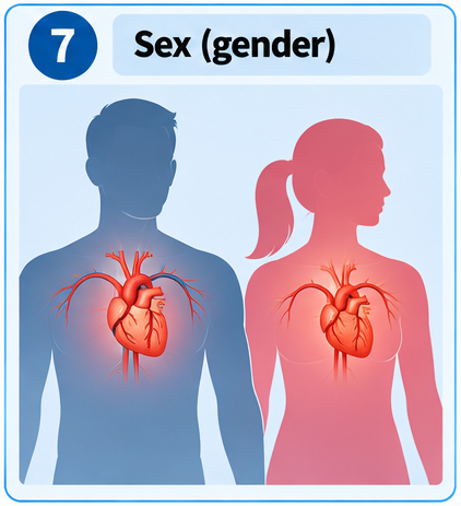
7. Sex: The risk of CHD differs between males and females and changes with age and other biological factors. Sex is a risk factor that cannot be modified.
These factors may act together. For example, a person with a genetic predisposition may further increase their risk through smoking, an unhealthy diet and physical inactivity. Having one or more risk factors does not mean that a person will inevitably develop CHD.
4.4 Prevention of Coronary Heart Disease
Although age, sex and genetic predisposition cannot be changed, lifestyle choices can help reduce the risk of CHD.
Balanced diet
A balanced diet provides the nutrients needed for normal body function while helping maintain a healthy body mass and cardiovascular health.
- Limit foods high in saturated fat and trans fat to reduce factors contributing to fatty deposits in artery walls.
- Choose sources of unsaturated fats, such as nuts, seeds and suitable vegetable oils.
- Eat plenty of fruit, vegetables and whole grains.
- Limit excessive salt intake to help control blood pressure.
- Maintain an appropriate energy intake to reduce the risk of excess body fat.
Regular physical activity
Regular physical activity helps maintain cardiovascular fitness and supports the regulation of body mass, blood pressure and blood lipid levels.
During exercise, heart rate increases to supply working muscles with oxygen and nutrients. With regular training, the cardiovascular system can adapt to physical demands, and the heart may pump blood more efficiently. A physically active lifestyle also helps reduce several risk factors associated with CHD.
Avoiding smoking
Avoiding tobacco smoking reduces exposure to chemicals that damage blood vessels, increase cardiovascular strain and promote blood clot formation. Avoiding second-hand smoke is also beneficial.
Managing stress and maintaining healthy habits
Managing persistent stress, getting adequate sleep and maintaining a physically active lifestyle can support cardiovascular health. Regular health checks can help identify risk factors such as high blood pressure or abnormal blood cholesterol levels.
No single lifestyle change eliminates the risk of CHD. Combining a balanced diet, regular exercise, avoiding smoking and appropriate medical care offers better protection than relying on one measure alone.
You have learned how the circulatory system transports substances, how the heart and blood vessels work, and how cardiovascular health is maintained. Now test whether you can explain the “why” and “how”, not just remember the facts.
Try to answer each question in your own words before revealing the explanation.
1. Why do humans need double circulation, and how does it benefit the body?
In double circulation, blood passes through the heart twice during one complete circuit of the body. The pulmonary circulation carries blood between the heart and lungs for gas exchange, while the systemic circulation carries blood between the heart and the rest of the body. This allows blood to be pumped to the lungs at lower pressure and to the body at higher pressure, ensuring efficient gas exchange and rapid delivery of oxygen to tissues.
2. Why is the muscular wall of the left ventricle thicker than that of the right ventricle?
The left ventricle pumps blood around the entire body, so it must generate high pressure to overcome the greater resistance of systemic circulation. The right ventricle pumps blood only to the nearby lungs, where lower pressure is sufficient. Therefore, the left ventricle has a thicker muscular wall than the right ventricle.
3. Why do arteries, veins and capillaries have different structures?
Each type of blood vessel is adapted to its function. Arteries have thick, muscular and elastic walls to withstand high blood pressure. Veins have thinner walls, wider lumens and valves to help return blood to the heart at low pressure. Capillaries have walls only one cell thick, providing a short diffusion distance for the exchange of substances between blood and tissues.
4. How do the different components of blood work together to maintain the body’s normal functioning?
Red blood cells transport oxygen using haemoglobin, while plasma transports dissolved substances such as nutrients, hormones, urea and carbon dioxide. White blood cells defend the body against pathogens through processes such as phagocytosis and antibody production. Platelets help blood clot when blood vessels are damaged, reducing blood loss and helping prevent pathogens from entering the body.
5. Why does heart rate increase during exercise, and why does it take time to return to its resting rate?
During exercise, muscles require more energy and their rate of respiration increases. The heart beats faster to deliver more oxygen and glucose to the muscles and remove carbon dioxide more rapidly. After vigorous exercise, heart rate remains elevated for some time to support recovery, including the transport of lactic acid to the liver and the supply of oxygen needed to deal with it.
6. How can narrowing of the coronary arteries affect the heart, and how can lifestyle choices reduce the risk?
Coronary arteries supply oxygenated blood to the heart muscle. Fatty deposits can narrow these arteries, reducing the supply of oxygen needed for aerobic respiration. This can cause chest pain and, if a coronary artery becomes severely blocked, a heart attack. A balanced diet, regular physical activity, avoiding smoking and managing other risk factors can reduce the likelihood of developing coronary heart disease.